What is Gas Gangrene?
Gas gangrene is caused by Clostridium perfringens, Cl. septicum, Cl. oedematiens and Cl. histolitycum. Gas gangrene usually develops with extensive crumbling of tissues (gunshot, torn, ragged-bruised wounds), often polluted with earth, scraps of clothing. The more tissues, especially muscles, are destroyed, the more favorable the conditions for the development of gas gangrene.
Causes of Gas Gangrene
The causative agents of gas gangrene are anaerobic microbes that constantly inhabit the intestines of domestic herbivores. They can be seeded from the skin and from the feces of practically healthy individuals. The nutrient medium is the dead muscles and other tissues that are in the wound. Reproduction of microbes occurs in an oxygen-free environment. Most of the anaerobic microbes in the process of life forms a gas. Anaerobic infection tends to spread rapidly, causing pronounced general intoxication of the body. Entrance gates of infection are most often traumatic tears of limbs, weakened wounds, much less often foreign bodies, injuries of the large intestine. Even a small wound can be complicated by an anaerobic infection. Anaerobic gangrene develops during the first days after the injury, at least – later.
Symptoms of Gas Gangrene
The clinical features of the flow of gas gangrene depend on the type of bacteria. So, Cl. perfringens is characterized by a toxic hemolytic, fibrinolytic, and necrotic course. Cl. septicum causes blood-serous swelling of tissues, while gas is released in small quantities and not always. The action of its toxins leads to hemolysis of red blood cells. The introduction of toxin Cl. Septicum in the experiment causes an immediate general reaction: a decrease in blood pressure, severe cardiac arrhythmias, depending on the damage to the heart muscle. Cl. oedematiens causes rapidly increasing tissue edema with the release of large amounts of gas; as well as other bacteria of this group, it releases hemolytic toxin. Cl. histolitycum is able to dissolve living tissue, melt muscles, connective tissue. After 10 to 12 hours of its impact, the soft tissues can be so destroyed that the bones become visible.
The clinical picture is characterized by local symptoms and general manifestations. In accordance with local symptoms, 4 forms of gas gangrene are distinguished:
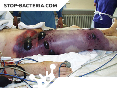
– emphysematous (classical) form. Local edema of tissues under the influence of microbes and their toxins passes into death with pronounced gas formation. The wound becomes dry, without signs of granulation, with extensive necrosis. On palpation of the wound area, the serum fluid and gas bubbles can be released from it. The skin around the wound becomes pale, cold, covers with brown spots. Crushed muscles are visible in the wound, which for several hours become dead, acquiring a gray-green shade. Sharply pain in the wound. Disappears pulse on peripheral arteries. With the destruction of the muscles appears corpse scent. The extremity gradually becomes brown, the sensitivity is lost and becomes dead throughout. At the same time pus is not formed;
– edematous toxic form. Initially, extensive edema is observed around the wound, and then it spreads far from the affected area. Gas formation is negligible. There is no purulent compartment. A bloody liquid in the color of meat is formed from the wound. The edema is growing literally before our eyes (if a thread is put around a limb, then after a few minutes the thread will start to “cut” into the skin). Muscles due to compression of edematous fluid become pale and protrude from the wound. Subcutaneous fatty tissue of a greenish shade of gelatinous-gelatinous type. The skin is sharply tense, shiny, cold to the touch. Disappears pulse and, quickly progressing, the death develops. Gas bubbles may be visible on x-rays. In this form of gas gangrene, wound gas is insignificant or absent altogether;
– phlegmonous form. This form of gas gangrene is less turbulent and is often limited to any area. In this form, it is even possible to distinguish between the depth of the process and highlight the deep and superficial forms of the lesion. Purulent discharge, with gas bubbles. Muscles are often pink in color, with areas of death. The inflammatory process often spreads through the intermuscular spaces. Typically, the local temperature of the skin does not decrease and the skin feels warm to the touch. As a rule, the pulse on the peripheral vessels is preserved. Spots on the skin are absent or not significant, as is the edema;
– putrid or putrid form. It usually develops very rapidly, accompanied by rapid decay. The process is distributed mainly in fiber, in the intermuscular spaces. A very rapid deceleration of the fascias of the muscles in the wound begins, and they acquire a dirty gray color. Discharge of putrefactive, with areas of dead tissue, with gas and a sharp putrid odor. Such changes are usually caused by a symbiosis of anaerobic and putrefactive bacteria. It should be noted that the causative agents of putrefactive infections have toxins that destroy proteins of any tissue, including the walls of blood vessels. Therefore, in this form, secondary erosive bleeding often occurs. If the first three forms are most often localized on the extremities, then the putrid form spreads near the rectum, mediastinum, etc.
Thus, the main local symptoms of gas gangrene are:
- puffiness;
- the presence of gas in soft tissues;
- muscle breakdown;
- the absence of symptoms characteristic of the inflammatory process.
Common symptoms.
The incubation period for anaerobic infection is short – 2 – 3 days. Rarely observed fulminant gas gangrene. Common symptoms of the disease:
- tachycardia;
- lowering blood pressure;
- excitement of the patient, talkativeness (sometimes, on the contrary, depressed mood);
- painful insomnia;
- body temperature from the very beginning of the disease increased, often above 38 – 39 ° C. Hyperthermia is a poor prognostic symptom;
- general intoxication, dehydration play a role;
- breathing is speeded up;
- pulse up to 120 – 140 beats per minute;
- erythrocyte hemolysis develops, which leads to rapidly developing anemia;
- hemoglobin level drops to 70 – 100 g / l;
- the number of red blood cells drops to 1 – 1.5. 1012 / l;
- there is a leukocytosis of up to 15 – 20. 109 / l with a shift of the leukocyte formula to the left due to an increase in stab neutrophils, with the advent of young forms, in the absence of eosinophils;
- the renal excretory function suffers sharply, oliguria develops, and then anuria. In severe cases, hematuria can occur.
The disease proceeds very rapidly, and if treatment is started at the wrong time, it quickly (within 2 to 3 days) causes death.
Diagnosis of Gas Gangrene
The diagnosis is made on the basis of a characteristic wound, general intoxication. The diagnosis is confirmed radiologically (determined by the “porosity” of muscle tissue) and microscopically (detection of clostridia in the wound discharge). The differential diagnosis is carried out with fascial gas-forming phlegmon (no muscle damage) and putrid (putrid) infection.
Treatment of Gas Gangrene
The main condition for the prevention of gas gangrene is timely and complete PCE of the wound with excision of non-viable tissues, as well as edges and bottom of the wound within healthy tissues. Such an operation should be performed under general anesthesia or conduction anesthesia. Experience shows that the hope for prophylactic actions of anti-gangrenous serums is unjustified. In addition, the introduction of serum often causes severe complications up to the development of anaphylactic shock. Since anaerobic bacteria are susceptible to antibiotics, for any extensive wound, even if subjected to adequate PEC, it is necessary to conduct both local and general therapy with broad-spectrum antibiotics.
Prevention of Gas Gangrene
If you suspect the development of gas gangrene, it is necessary to combine active surgical treatment with vigorous general measures. It is necessary to conduct a wide dissection of all suspicious areas, excise all non-viable tissue. Wide parallel (Lompast) incisions should cut the fascia and soft tissue to the full depth. Proper drainage should ensure the outflow of discharge from the wound. It should be emphasized that wounds must remain open. An introduction to the bottom and edges of the wounds of broad-spectrum antibiotics is necessary. When confirming the diagnosis and spreading gangrene, immediate amputation or exarticulation of the limb is necessary. The wound after amputation cannot be sutured. As a supplement to the operation, but not instead of it, hyperbaric oxygenation is shown. For this purpose, the patient is placed in a chamber with elevated pressure (up to 3 atmospheres), on the first day at least 3 times 2 to 2 1/2 hours. On subsequent days, the sessions can be held once a day.
In addition, immediately after the detection of gangrene, intensive infusion therapy with the introduction of albumin, plasma, electrolyte solutions and proteins is necessary. Patients with anemia are transfused with freshly prepared single-group whole blood or erythrocyte mass. At the same time, high doses of antibiotics are administered intravenously or intraarterially.
Anti-gangrenous serum (monovalent when the causative agent is detected, and polyvalent if not detected) is administered intravenously at a dose of 150000 AE. The serum is dissolved in an isotonic solution of sodium chloride and heated to 36 – 37 ° C.
Patients with gas gangrene should be isolated. They should have a separate nursing post. All underwear, tools must be specially processed. It is important to remember that the vegetative forms of bacteria die by boiling, and their spores retain their vital functions and die only when fractional (repeated) boiling. It is better if the instruments are subjected to air sterilization (in a dry-heat oven) at t 1500С, or sterilization in a steam sterilizer under pressure of 2 – 2 1/2 atmospheres.
Health care workers who care for the sick must observe personal hygiene. Dressings, treatment of the oral cavity, skin should be made in rubber gloves, which should be disinfected regularly (chloramine, carbolic acid, lysol, etc.) after each dressing. All dressings should be burnt immediately after dressing.