What is Peripheral Lymph Node Tuberculosis?
Tuberculosis of peripheral lymph nodes in adults according to the clinical picture does not differ from that in children. In the absence of HIV infection, it can be considered as a variant of primary tuberculosis. However, some details need to be noted.
Causes of Peripheral Lymph Node Tuberculosis
Mycobacterium tuberculosis (MBT) – facultative intracellular parasites.
Mycobacterium tuberculosis (MBT) belong to the family of bacteria Micobacteriacae, order Actinomycetalis, genus Mycobacterium. The genus Mycobacterium has over 100 species, most of which are saprophytic microorganisms, widely distributed in the environment.
Etymologically, the word “mycobacterium” comes from the Greek words myces – mushroom and bacterium, bactron – stick, twig. The component of the name “mushroom” is due to the tendency of these microorganisms to form filamentous and branching mold-like forms.
From the standpoint of clinical medicine, mycobacterium tuberculosis, discovered by the German scientist Robert Koch, is the most important type of actinomycetes, which are combined in a complex including M. tuberculosis (MBT); M. bovis and its variant BCG (Bacillus Calmette-Guerin); M. africanum and M. microti. This group of mycobacteria has a pronounced genetic similarity.
M. microti is not considered pathogenic to humans, but causes a disease in mice that resembles tuberculosis. BCG culture is not pathogenic to humans. Mycobacterium tuberculosis (MBT) is up to 95% of cases the cause of human tuberculosis, depending on the territory of residence. At the same time, M. bovis and M. africanum cause a disease in humans that is not clinically different from classical tuberculosis.
Mycobacteria, not included in the M. tuberculosis complex, can cause mycobacteriosis. Such mycobacteria are combined into complexes, the most important of which are: M. avium, M. fortinatum and M. terrae, M. leprae, M. ulcerance.
The materials on tuberculosis presented below are relevant only to the disease caused by M. tuberculosis (MBT) – Koch bacteria (CD), typus humanus.
The natural reservoir of tuberculous mycobacteria is humans, domestic and wild animals, birds.
MBT externally are thin curved sticks resistant to acids, alkalis and drying. The outer shell of the bacteria contains complex waxes and glycolipids.
MBT can multiply both in macrophages and outside cells.
MBTs multiply relatively slowly. Reproduction occurs mainly through simple cell division. On enriched nutrient media, MBT multiply with a doubling period of 18 to 24 hours. For growth in a culture of mycobacterium tuberculosis, obtained under clinical conditions, it takes 4 to 6 weeks.
The genetic structure of the Office is established. The nucleotide sequence of the Office can be found in international data banks. The nucleotide sequence of the Office (strain H37Rv) has 4,411,529 b.p.
The Office does not possess independent movement. The temperature limits of growth are between 29 and 42 ° C (optimal – 37-38 ° C). MBTs are resistant to physical and chemical agents; they remain viable at very low temperatures, and can withstand elevations up to 80 ° C for 5 minutes.
In the external environment, mycobacterium tuberculosis is quite stable. In water, it can last up to 150 days. Dried mycobacteria cause tuberculosis in guinea pigs after 1-1.5 years, lyophilized and frozen are viable up to 30 years.
With intense exposure to the sun and at high ambient temperatures, the viability of the office environment decreases dramatically; on the contrary, in darkness and dampness, their survival is very significant. Outside of a living organism, they remain viable for many months, especially in dark, damp rooms.
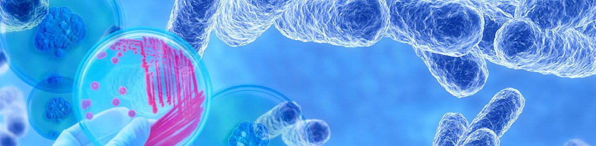
MBTs are detected using a unique staining property (acid resistance) that distinguishes them from many other infectious agents. Ziel (Ziehl) and Nelsen (Neelsen) in 1883 developed a special contrasting method for staining MBT, based on the property of acid resistance. Unlike non-acid-resistant bacteria, tuberculous mycobacteria are colored red, do not discolor when exposed to an acid solution, and are clearly visible on a blue background under microscopy. The Ziel-Nielsen method is still one of the main methods for staining MBT under microscopy. More sensitive than the acid-resistant staining method is MBT auramine staining followed by fluorescence microscopy.