
What is Toxoplasmosis?
Toxoplasmosis is a parasitic disease characterized by damage to the nervous system, eyes, skeletal muscles and heart muscles, as well as an increase in lymph nodes, liver and spleen. Prone to chronic course.
The prevalence of toxoplasmosis in the world is incredibly high, mainly due to African countries, as well as Latin and South America, in which the infection rate of the population reaches 90%. Rates in Europe and North America are lower – 25-50% of the population.
Causes of Toxoplasmosis
The causative agent of toxoplasmosis, Toxoplasma gondii, belongs to the protozoa type (Protozoa), the class of sporozoans (Sporozoa), the coccidia order (Coccidia). Toxoplasmas are mobile and have the form of an arc, arches or resemble an orange slice. There are also oval and rounded shapes. The type of movement in Toxoplasma sliding.
Pathogenesis during Toxoplasmosis
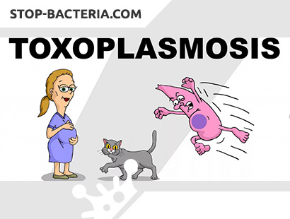
The life cycle of Toxoplasma includes the stages of sexual and asexual reproduction. Toxoplasmosis is common in many countries. A person becomes infected from domestic animals, most often from cats, which are the ultimate owner of the parasite. In their body, sexual reproduction of the pathogen occurs. Sick animals with toxoplasmosis produce parasites with urine, saliva, and milk. Man is an intermediate owner. Infection occurs by alimentary (most often), drip, through damaged skin and mucous membranes, transmissible (by arthropod bite) by. Perhaps intrauterine infection when the parasite penetrates from the mother to the fetus through the placenta. Toxoplasma infected from 50 to 80% of the adult population.
Ways to Toxoplasmosis
Infection occurs when eating meat products and eggs that have not undergone sufficient heat treatment. The possibility of infection if the pathogen enters the mucous membranes and damaged skin, transmissive, etc., is not excluded. There is also an intrauterine infection.
Factors that can contribute to the parasite in the body and increase the risk of toxoplasmosis:
- Touching with dirty hands to the mouth after contact with the ground, after cleaning the cat litter, or any other contact with the cat excrement.
- Eat raw or undercooked meat, especially pork, lamb or venison.
- Touch the mouth after contact with raw or uncooked / stewed meat.
- Organ transplantation or blood transfusion (very rarely).
- If a woman is pregnant and she has become infected with toxoplasmosis, the infection can be transmitted from her to the child, which can lead to serious consequences.
In humans, toxoplasma multiplies in the intestine, spread by lymphogenous and hematogenous. The phase of lymphogenous drift (lymph nodes are enlarged and inflamed) is replaced by hematogenous dissimination. The stage of finding the parasite in the blood is short (several days). Once in the internal organs, Toxoplasma causes inflammation in them. The nervous system, the retina, the liver, and the myocardium are particularly often affected. In these organs, toxoplasmas are intracellular and extracellular. Clusters of parasites are called pseudocysts. Toxoplasma can form cysts in tissues, causing a state of latent infection. The parasite is activated when conditions are unfavorable to the microorganism and its immunological reactivity decreases. In the pathogenesis of the toxoplasmosis of the nervous system, focal inflammatory phenomena (necrotizing encephalitis), dyscirculatory disorders associated with vasculitis, and obstruction of the cerebrospinal fluid, leading to hydrocephalus and microcephaly, are important.
The most gross morphological changes in the nervous system are observed in children. At macroscopic examination revealed expansion of the ventricles with periventricular zone of necrosis. Scars are found, replacing areas of necrosis, obliteration of the interventricular opening and lateral aperture of the IV ventricle. Hydrocephalus can be expressed, leading to thinning and deformation of the substance of the hemispheres.
Morphological manifestations of toxoplasmosis of the brain in adults are diverse. Microscopic examination is most characteristic of miliary granulomas scattered throughout the brain and spinal cord, consisting of large epithelioid cells, lymphocytes, monocytes, and sometimes eosinophils. Granulomas contain numerous parasites, surrounded by a zone of edema with necrotic foci caused by vasculitis. Typical for toxoplasmosis is the calcification of small foci. In the presence of toxoplasm seroproductive leptomeningitis occurs in the subarachnoid space.
Most infected have no clinical manifestations of the disease. In some patients, sluggish chronic forms are observed and extremely rarely acute, with a severe course of the disease. With intrauterine infection in the first months of pregnancy, miscarriage and fetal death often occur. The possibility of fetal deformities and the birth of children with developmental defects is not excluded. If the infection occurred in late pregnancy, a child is born with generalized toxoplasmosis.
Symptoms of Toxoplasmosis
Toxoplasmosis affects people of any age, but most often children. Allocate acquired and congenital toxoplasmosis.
Acquired Toxoplasmosis. The incubation period is from 3 to 14 days. The prodromal period with general malaise, muscle and joint pain – usually several weeks, sometimes months. The acute stage of the disease is manifested by fever, chills, lymphadenopathy. A generalized maculopapular rash appears, absent only on the soles, palms, scalp. Along with the general signs of an infectious disease, there is a clinical picture of damage to various organs: myocarditis, pneumonia, focal necrotic nephritis, hepatitis. Damage to the nervous system is manifested by meningitis, encephalitis, meningoencephalitis, encephalomyelitis. Radiculoneuric and low-symptom forms are rarely observed (the latter can only be detected by serological reactions).
The most typical form of toxoplasmosis is meningoencephalitis, in the clinical picture of which there are cerebral and meningeal symptoms, paresis of the limbs, tonic-clonic seizures, oculomotor (diplopia) and coordinating disorders. Sometimes single or multiple toxoplasmosis abscesses develop in the brain. Characteristic disorders of consciousness, lethargy, loss of memory and orientation in space. Leukocytosis with a shift of the formula to the left, an increase in ESR, in the cerebrospinal fluid – lymphocytic pleocytosis, a moderate increase in protein content are found in the blood.
Acute, chronic and latent forms are distinguished, with the latter being divided into primary latent, without clinical manifestations, and secondary latent, arising after the acute form or chronic relapse.
Acute toxoplasmosis is characterized by a sudden onset, fever, general intoxication. Patients have lymphadenopathy, maculopapular rash, enlarged liver, spleen. Signs of encephalitis, meningoencephalitis, and myocarditis may develop.
Encephalitic, typhoid-like and mixed forms of acute toxoplasmosis are distinguished depending on the prevailing syndrome.
After the process subsides, acute toxoplasmosis becomes secondary-chronic or, less commonly, secondary-latent.
Chronic toxoplasmosis can develop both a primary chronic and a secondary chronic disease. It is characterized by a torpid, prolonged course in which periods of exacerbation and remission are observed. The main clinical symptoms are intoxication, subfebrile condition, myalgia, arthralgia. Patients have irritability, memory loss, neurotic reactions. A common symptom is generalized lymphadenopathy. As a result of mesadenitis, aching pains and bloating, constipation, nausea occur. Among the important symptoms of the disease are specific myositis (in the thickness of the muscles you can sometimes feel compaction and even calcifications) and myocarditis. Many patients show signs of vegetative-vascular dystonia, endocrine disorders (menstrual disorders, impotence, secondary adrenal insufficiency, etc.). Often there is eye damage in the form of chorioretinitis, retinitis, uveitis. In the peripheral blood – leukopenia, neutropenia, relative lymphocytosis, tendency to eosinophilia. ESR is within normal limits.
However, the predominant form of the course of the acquired disease is latent toxoplasmosis. It often has a primary latent and, much less often, secondary latent character. Latent toxoplasmosis is diagnosed only by serological examination. Latent and chronic forms of the disease can go into a severe generalized course, which occurs with HIV infection and other conditions leading to immunodeficiency. The most common cases of HIV / AIDS in patients with meningoencephalitis and brain abscesses caused by toxoplasma. Computed tomography is of great importance in the diagnosis. The lungs, heart, myocardium and other organs are also involved in the process. Toxoplasmosis is one of the causes of death of HIV / AIDS patients.
Congenital toxoplasmosis. When the mother becomes ill with toxoplasmosis in the first half of pregnancy, the fetus, as a rule, dies due to developmental defects incompatible with life. When the mother is infected in the second half of pregnancy, the baby is born with severe brain damage. The acute stage of the disease occurs in utero, a child is born with actively ongoing meningoencephalitis or its consequences. The neurological manifestations of meningoencephalitis are diverse: polymorphic seizures, clonic-tonic convulsions, spastic paresis, tremors, myoclonus, paresis of the eye and facial muscles, nystagmus, muscle contractures, meningeal phenomena. Sometimes there are symptoms of spinal cord lesions.
Congenital toxoplasmosis is characterized by a triad of symptoms: hydrocephalus, chorioretinitis and intracerebral calcifications. With hydrocephalus, the size of the head increases, the bones of the skull become thinner, fontanelles are strained. Hydrocephalus is usually accompanied by microphthalmia. If hydrocephalus develops before the birth of a child, then in childbirth it is necessary to resort to craniotomy. However, in many cases, an increase in head volume is not detected and hydrocephalus is detected only with pneumoencephalography. Chorioretinitis is often bilateral, focal, including the macular region. There are also iritis, uveitis, cataracts, primary or secondary atrophy of the optic nerves. Intracerebral calcifications 1-3 cm in diameter are located in the cortex and basal ganglia and are detected on craniograms of CT and MRI of the brain.
Children with congenital toxoplasmosis lag behind in mental development up to oligophrenia. They also have a variety of psychotic states (depression, psychomotor agitation, hallucinations, catatonia). Sometimes in children with congenital toxoplasmosis jaundice, enlarged liver and spleen are found. The temperature usually remains normal. Lumbar puncture usually has normal cerebrospinal fluid pressure. High protein content and moderate mononuclear pleocytosis, sometimes xantochromia, are noted.
Complications of congenital toxoplasmosis are the result of damage to the brain and eyes, leading to exhaustion, paralysis, mental retardation, blindness. Joining a secondary infection causes the development of purulent meningoencephalitis and pyocephaly. Death occurs from progressive brain damage.
The course of toxoplasmosis
It was previously believed that congenital toxoplasmosis leads to death during the first years of a child’s life. At present, stabilization of the infection and even full recovery with residual effects are possible, the severity of which depends on the degree of damage to the central nervous system (calcifications, choreoretinitis, epileptic syndrome, mental retardation, etc.). In adults, along with the acute course, a subacute or even chronic development of the disease is often noted. Often acquired toxoplasmosis, especially in adults, proceeds without clinical symptoms expressed to one degree or another (inapparent form).
Diagnosis of Toxoplasmosis
The diagnosis of toxoplasmosis is based on a set of indicators that include clinical data and laboratory confirmation results. The epidemiological prerequisites are of relative importance, such as contact with cats, the conditions for communicating with them, especially eating habits (eating raw, half-baked meat, a sample of raw minced meat, unwashed vegetables, fruits), personal hygiene skills, profession, etc.
The extreme variety of clinical manifestations, the absence of symptoms characteristic of toxoplasmosis only, complicates the clinical diagnosis and, in certain cases, only suggests a diagnosis that is formed during a full-fledged differential diagnosis and in the analysis of laboratory results.
Laboratory methods for the diagnosis of toxoplasmosis are divided into two groups: parasitological and immunological.
Parasitological methods are based on the possibility of detecting a pathogen or isolating it during infection of susceptible animals – a bioassay.
These include direct microscopy of smears – fingerprints of affected organs (tonsils, biopsy specimen of the lymph nodes, brain, internal organs of dead embryos or fetuses) or smears from a sediment of cerebrospinal fluid, blood stained according to Romanovsky-Giemsa. You can examine the histological preparations of these organs.
Unfortunately, the technical difficulties of detecting post mortum, as well as the rare cases of detecting toxoplasmas in the blood, cerebrospinal fluid make it difficult to use these methods.
A biological test on white mice, hamsters infected with the test material and subsequent 5-6 blind passages requires special conditions for keeping animals, laboratories with a special mode of operation and is used only for scientific purposes.
In general practice, mainly immunological methods are used, which include serological reactions and an intradermal test. These methods, quite specific and sensitive, determine, first of all, the state of infection, and seroreaction – and the incidence. These concepts are ambiguous, because the incidence is many times lower than infection.
Of the methods of serological diagnostics, the complement binding reaction (CSC), the indirect immunofluorescence reaction (RNIF) and enzyme-linked immunosorbent assay (ELISA) are used. The diagnosis is confirmed by a significant increase in antibody titer – the dynamics of titers in paired serum taken with an interval of 2-4 weeks.
CSC becomes positive from the 2nd week after infection and reaches the highest titers – 1:16 – 1: 320 reaches after 2-4 months. After 1-3 years, it can become negative or persist in low titers (1: 5, 1:10), which do not have independent significance.
RNIF becomes positive from the 1st week of infection and reaches its maximum level (1: 1280 – 1: 5000) at 2-4 months. In low titers of 1:10 – 1: 40, it can last 15-20 years.
ELISA in accordance with the orientation to the international standard of WHO is the most objective method. A positive reaction is indicated by optical indicators of more than 1.5, in immunofermet units – more than 60; in international – more than 125, in antibody titers – 1: 1600 and more.
Diagnosis of congenital toxoplasmosis in a child begins with an obstetric history of the mother, an epidemiological history and indicators of serological reactions. Specialist consultations are required for differentiation with herpetic, cytomegalovirus, listeriosis, chlamydial infections, as well as x-ray of the skull and examination at the medical center.
It should be recalled that 20-30% of women have antibodies – these are healthy carriers of antibodies. They do not require treatment. 70 – 80% who showed negative seroreactions, are at risk and need re-examination.
During the first year of a child’s life, it is necessary to conduct parallel serological examinations of the mother and child in dynamics.
Positive reactions in the mother and the child in the first three months of the child’s life do not give reason for the diagnosis of “toxoplasmosis” in the child, because specific antibodies in the IgG composition are transmitted to the child transplacentally. To confirm the diagnosis of congenital toxoplasmosis in the newborn, the Remington test is a variant of the RIF with the determination of IgM that do not pass through the placenta. Their detection indicates infection of the fetus.
Toxoplasmosis Treatment
In acute toxoplasmosis, chemotherapeutic drugs are used.
Delagil (0.5 g 2 times a day) in combination with sulfonamides (0.5 g 2 times a day) for 10 days. Fansidar is prescribed in the amount of 5 tablets per course: 1 tablet. after 2 days or in the form of intramuscular injections, 1 ampoule of 2.5 mg once every 2 days in the amount of 5 injections. One or two courses of treatment are carried out.
Of the antibiotics prescribed: lincomycin hydrochloride (0.5 g 2 times a day); metacyclin hydrochloride (0.3 g 2 times a day) for 5-7 days in combination with sulfonamides; rovamycin.
Treatment of chronic toxoplasmosis is many times more difficult than acute, since chemotherapy does not have a significant effect. The main place is occupied by hyposensitizing and immunomodulating therapy. The complex of treatment includes vitamins, desensitizing agents, lidase, cerebrolysin, etc.
There is evidence of a positive effect of levamisole in the treatment of chronic toxoplasmosis. Levamisole is prescribed at 150 mg for 3 days in a row with 1 week intervals between cycles, a total of 2 to 3 cycles.
Toxoplasmosis Prevention
Prevention of toxoplasma infection consists in eating only well-thermally processed meat and meat products, cleanly washed vegetables, fruits and berries. In the process of cooking, it is forbidden to taste raw minced meat. It is necessary to wash your hands thoroughly after processing raw meat products, working in the garden, garden, children after playing in the playground and, especially, in the sandbox. It is necessary to carefully observe the sanitary and hygienic rules for keeping pets in the apartment, not forgetting to wash their hands after contact with them.
When pregnancy occurs, every woman should be examined for toxoplasmosis in a antenatal clinic. If a pregnant woman manifests clinical manifestations of toxoplasmosis, as well as the detection of antibodies to IgM class toxoplasma, the question of the need for treatment or termination of pregnancy should be addressed.